 Exosome RNA Exosome RNA Research & Industry News

Extracellular vesicles, including exosomes, are tiny membrane-bound particles released by cells into blood and other body fluids. They carry RNA, DNA, and proteins that reflect the state of the cells they come from, making them powerful tools for liquid biopsy applications in cancer, infectious disease, and neurological disorders.
A team led by researchers at Tulane University School of Medicine describes a new approach that simplifies how these vesicles are analyzed directly in patient blood. Traditionally, studying vesicle-associated biomarkers required isolating EVs first, a process that is slow, labor-intensive, and difficult to scale for clinical use.
The method outlined in this work avoids isolation altogether. Instead, antibodies are used to capture extracellular vesicles directly from blood samples. Reagent-loaded liposomes are then added, which fuse with the vesicle membranes and deliver detection components straight into the vesicles. Once inside, CRISPR-based molecular tools detect specific nucleic acid targets with high sensitivity.
Applications of liposome–EV fusion systems in diagnostics,
therapeutics, regenerative medicine and immune modulation
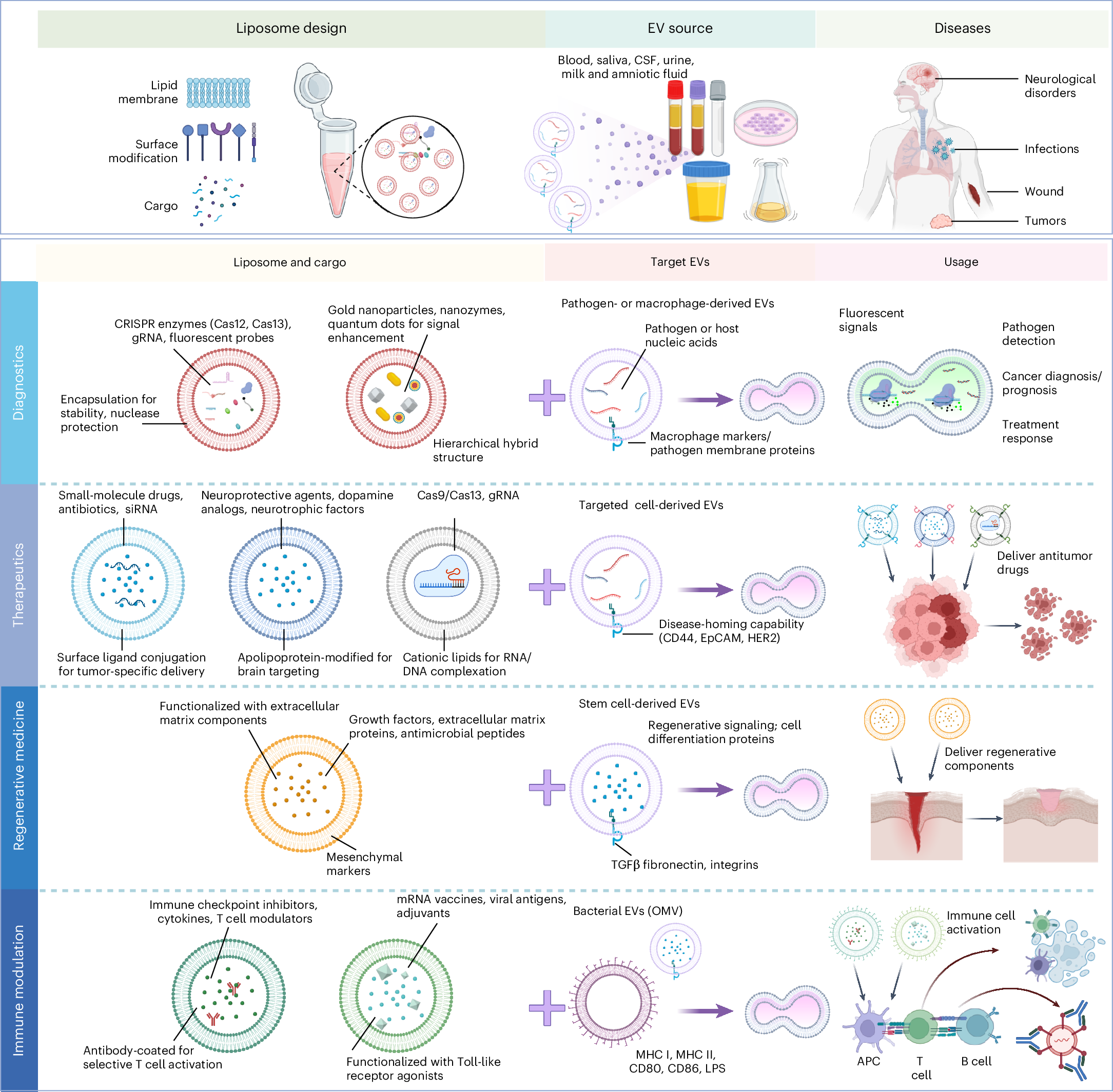
This schematic illustrates the design of liposomes, their integration with EVs from various biological sources (for example, blood, saliva, cerebrospinal fluid, urine, milk and amniotic fluid) and their applications across multiple disease contexts, including neurological disorders, infections, wounds and cancer. The top illustration shows liposome features (lipid membrane, surface modifications and cargo loading), EV sources and related diseases. The bottom illustration is divided into four application areas: diagnostics, liposomes encapsulating CRISPR systems or nanomaterials that detect pathogen- or host-derived NA from EVs for disease detection and monitoring; therapeutics, drug-loaded liposomes targeting disease-associated EVs for tumor- or brain-specific delivery; regenerative medicine, bioactive cargo and surface markers enhancing fusion with stem cell-derived EVs for tissue repair; immune modulation, liposomes functionalized with immunoregulatory agents or antigens engaging bacterial EVs to activate immune cells, offering platforms for immunotherapy or vaccination. APC, antigen-presenting cell; CSF, cerebrospinal fluid; LPS, lipopolysaccharide; siRNA, small interfering RNA.
This liposome–EV fusion strategy was initially developed to detect viral RNA inside vesicles for infectious disease diagnosis. Since then, many groups have adapted it to measure mRNA, microRNA, DNA mutations, and even surface proteins from tumor-derived vesicles. Because the vesicle membrane protects nucleic acids from degradation, the assay can work directly in complex samples like plasma.
By combining direct vesicle capture, membrane fusion, and programmable CRISPR detection, the approach offers a flexible platform for both research and clinical diagnostics. It opens the door to faster, less invasive tests that better reflect what is happening inside diseased tissues, without the bottlenecks of traditional vesicle isolation.


