 Exosome RNA Exosome RNA Research & Industry News

Understanding tiny particles like nanoparticles and extracellular vesicles (EVs) is becoming increasingly important in fields like drug delivery, diagnostics, and environmental science. These particles are incredibly small—thousands of times smaller than a human cell—and even particles that look similar can behave very differently. That’s why scientists need tools that can analyze them one at a time, rather than in bulk.
In this work at Vanderbilt University, researchers developed a new platform called Interferometric Electrohydrodynamic Tweezers (IET). This technology is designed to trap and analyze single nanoparticles quickly and efficiently.
Working principle of the Interferometric Electrohydrodynamic Tweezers (IET) system
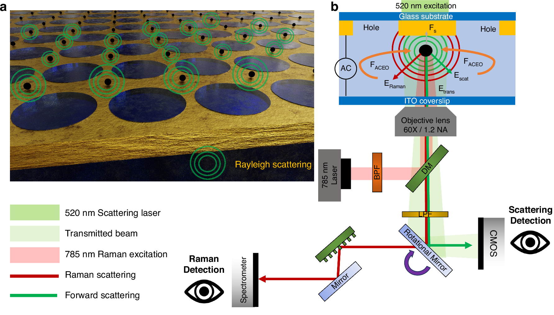
a Schematic view of the IET system capable of rapid parallel trapping of multiple particles and label-free interferometric imaging. b Illustration of the working principle and experimental setup of the IET system. Nanoparticles are rapidly transported by in-plane AC electro-osmotic (ACEO) flow and trapped at the nearest stagnation zones. Forces acting on the particles include FACEO, the drag force from ACEO flow, and Fs, the particle surface interaction force. Optical signals involved are Escat, the scattered electric field from the particle; ERaman, the Raman signal from the particle; and Etrans, the transmitted light after passing through the thin gold film. Optical components include: DM the dichroic mirror, BPF the band-pass filter, and LPF the long pass filter
Traditional methods for studying nanoparticles often come with trade-offs. Some are slow, taking minutes to analyze just one particle. Others can damage or permanently trap particles, making repeated measurements difficult. Many also struggle to work when particle concentrations are low, which is common in real biological samples.
The IET platform solves many of these challenges by combining several advanced techniques into one system. It uses electrical forces to gently trap individual particles in solution, while simultaneously analyzing them using interferometric scattering imaging (to determine size and shape) and Raman spectroscopy (to identify chemical composition).
What makes this approach particularly powerful is speed and efficiency. Instead of taking minutes per particle, IET can trap and analyze nanoparticles in just seconds. This allows researchers to examine many more particles in a shorter time, providing a clearer picture of particle diversity, also known as heterogeneity.
The researchers demonstrated the system using both synthetic polymer nanoparticles and extracellular vesicles, which are naturally released by cells and play key roles in cell communication. Because EVs are being explored as biomarkers and drug delivery vehicles, having a fast and detailed way to characterize them is especially valuable.
Another important advantage is that IET works well even when particles are present at very low concentrations. This is critical for real-world biological samples, where EVs and other nanoparticles may be scarce.
Overall, this technology opens new possibilities for studying nanoscale particles in a more detailed and efficient way. Applications could range from improving drug delivery systems to better understanding how extracellular vesicles function in health and disease, and even monitoring environmental nanoparticles.


