 Exosome RNA Exosome RNA Research & Industry News

Isolating tiny biological particles from blood, saliva, and other body fluids is a major challenge in modern biomedical research. These particles, such as viruses and small extracellular vesicles (sEVs), are incredibly small, yet they play outsized roles in infection, immune signaling, and disease progression. Improving how researchers isolate them can directly impact diagnostics, drug delivery, and personalized medicine.
A team led by researchers at Duke University has developed a detailed and standardized protocol using sound waves to reliably separate these nanoscale particles from biofluids. Their work provides practical guidance that could help many laboratories adopt acoustic separation methods more easily and consistently.
Summary of the fabrication and sample processing for the acoustic
nanoparticle-separation device, corresponding to the protocol steps
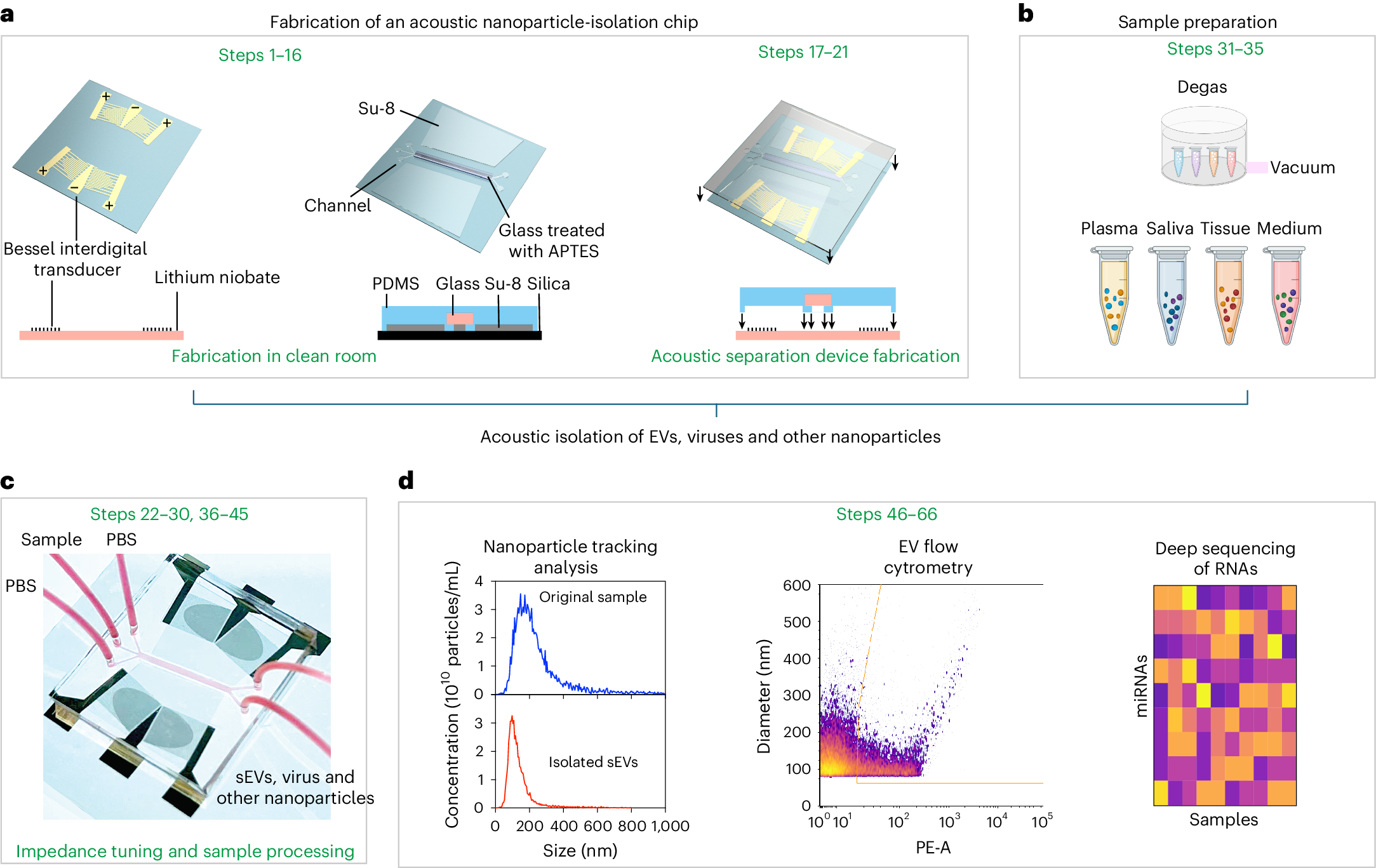
a, A schematic illustration of the fabrication process for the acoustic separation device. The device integrates microfluidic channels and a Bessel interdigital transducer to generate standing surface acoustic waves. b, A diagram summarizing the preparation of various sources of samples before acoustic separation. c, An illustration of the tuning and impedance matching that are required before sample processing. Device image reproduced with permission from ref. 46, ACS. d, An overview of the analytical results steps.
Why small extracellular vesicles are important
Small extracellular vesicles, sometimes called exosomes, are membrane-bound particles released by cells into body fluids. They carry proteins, RNA, and other molecules that reflect the state of their cell of origin. Because of this, extracellular vesicles are increasingly studied as biomarkers for cancer, infectious diseases, and inflammatory conditions. They are also being explored as natural delivery vehicles for drugs and genetic therapies.
Viruses are similar in size to extracellular vesicles, which makes separating one from the other especially challenging. Traditional isolation techniques—such as ultracentrifugation or filtration—can be slow, labor-intensive, and prone to variability between laboratories.
How acoustic separation works
Acoustic separation uses precisely controlled sound waves to move particles within a microfluidic channel. When an acoustic field is applied, particles experience forces based on their size, density, and mechanical properties. This allows researchers to gently guide viruses and extracellular vesicles into specific regions where they can be collected.
One major advantage of this approach is that it does not rely on chemical labels or harsh processing steps, helping preserve the integrity of sensitive nanoscale particles. It also allows relatively high throughput, making it suitable for routine laboratory use.
What makes this protocol important
While acoustic techniques have shown promise before, their wider adoption has been limited by a lack of standardized fabrication and validation methods. In this work, the authors address that gap by providing a comprehensive, step-by-step protocol that covers:
- Design and fabrication of the acoustic separation device
- Optimization of the acoustic field for nanoscale particles
- Preparation of plasma and saliva samples
- Validation of performance across different operators
- Data collection and analysis to assess particle purity and integrity
By demonstrating reproducibility and performance consistency, the protocol lowers the barrier for other laboratories to implement acoustic isolation in their own workflows.
Broader impact across biology and medicine
Standardized acoustic isolation of viruses and extracellular vesicles could accelerate discoveries in virology, immunology, and cell-to-cell communication. Reliable access to high-quality extracellular vesicles may improve liquid biopsy development, support regenerative medicine strategies, and enable new therapeutic delivery platforms.
Beyond medicine, the authors note potential applications in materials science and bioengineering, where controlled manipulation of nanoscale particles is increasingly important.
As interest in extracellular vesicles and nanoscale diagnostics continues to grow, robust and reproducible methods like this acoustic protocol will be critical for translating basic research into real-world clinical and technological advances.
Xia J, Lu B, Yang S, Ghosh A, Chang C, Hakim A, Mai JDH, Chen Y, Wong DTW, Nolan JP, Laurent LC, Dao M, Sadovsky Y, Lee LP, Suresh S, Huang TJ. (2026) Acoustic separation and isolation of viruses, small extracellular vesicles and other nanoscale bioparticles. Nat Protoc [Epub ahead of print]. [article]


