 Exosome RNA Exosome RNA Research & Industry News

Researchers are constantly searching for new ways to stop cancer from spreading, and a team led by researchers a Southern Medical University has taken an exciting step forward. They’ve developed a nanotechnology-based therapy that not only targets tumors directly but also blocks the tiny messengers that help cancer cells spread throughout the body.
These messengers are called tumor extracellular vesicles (TEVs), microscopic packages released by cancer cells that carry signals to nearby and distant tissues. TEVs play a key role in metastasis, the process by which cancer spreads to other parts of the body, and are known to influence immune evasion, tissue remodeling, and even drug resistance.
The team engineered lipid-coated nanoparticles using palmitic acid that can be taken up by tumor cells and incorporated into their TEVs. What’s novel is that these nanoparticles serve a dual purpose: they track the TEVs and disable them. Once the nanoparticles are inside the cells and vesicles, the researchers shine near-infrared light on the tumor site, which activates the particles to release reactive oxygen species (ROS), toxic molecules that damage the cancer cells and disrupt the TEVs.
This light-triggered photodynamic therapy caused a double blow: it shrank the primary tumor and simultaneously shut down the communication network that allows cancer to spread. The approach was tested in multiple tumor models in female mice and showed significant inhibition of both tumor growth and metastasis.
Concurrent inhibition of tumor growth and metastasis by NP3
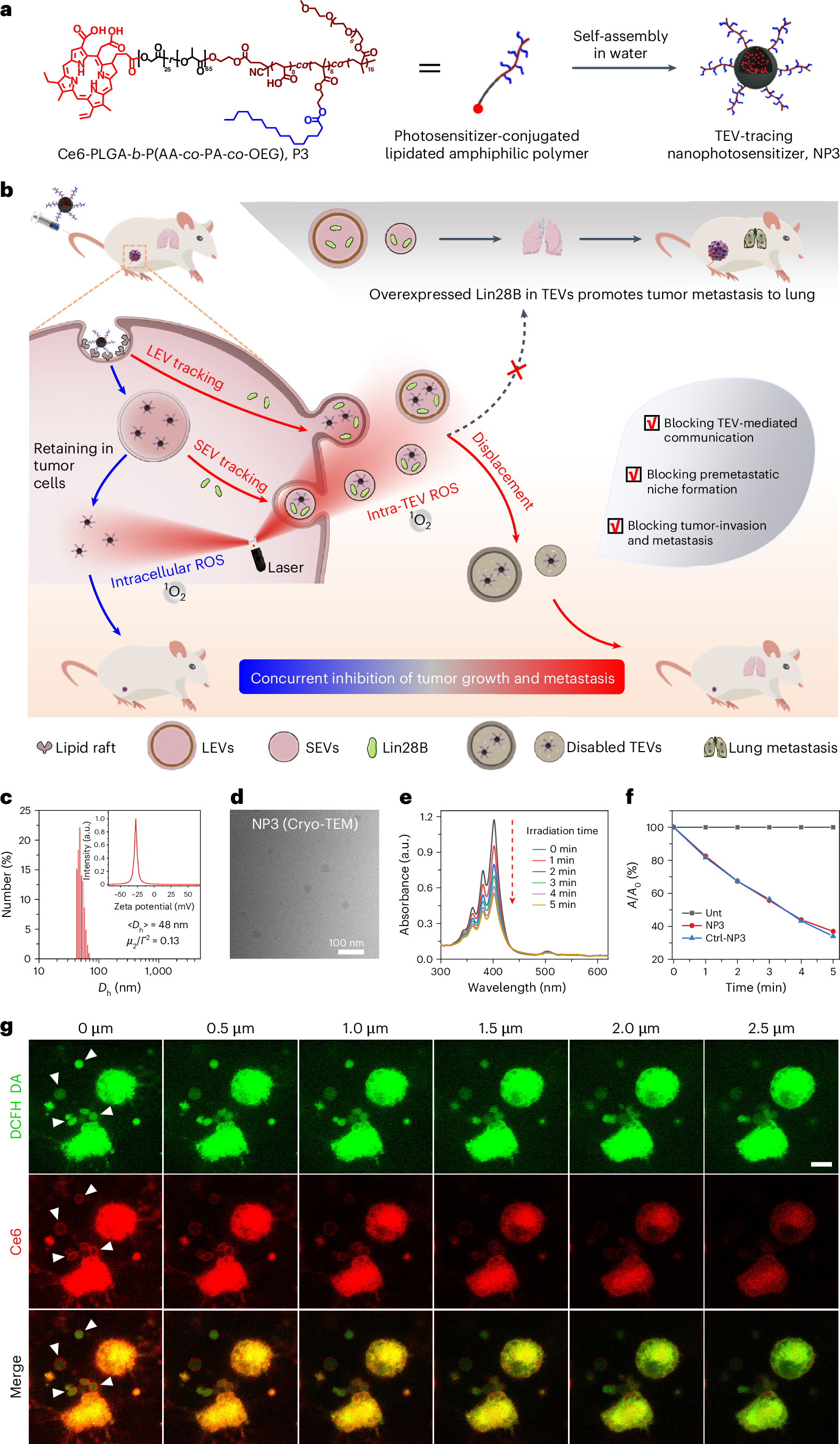
a, The molecular structure and preparation of NP3. The lipidated amphiphilic polymer is composed of a hydrophobic PLGA chain segment and hydrophilic random copolymer chain segment containing AA, OEG and PA. The photosensitizer (Ce6) is covalently attached to the end of the polymer chain. PA moieties exposed to the surface of the nanoparticle after self-assembly in water, which provide functions of molecular interaction with tumor cells. b, Therapeutic mechanism illustration of concurrent inhibition of tumor growth and metastasis by NP3 tracing and disabling TEVs. After uptake by tumor cells, NP3 distributes both in cells (retained in tumor cells) and in TEVs (LEVs and SEVs). By irradiating the tumor with a 660-nm laser, NP3 can not only kill the primary tumor cells by producing intracellular ROS but also efficiently destroy the TEVs by producing intra-TEV ROS. The latter function blocks TEV-mediated communications, premetastatic niche formation and tumor invasion and metastasis by destroying the Lin28B-overexpressing TEVs. c–g, Intracellular and intra-TEV ROS generation of NP3. c, Size distribution and zeta potential (inset) of NP3. d, Cryo-TEM image of NP3. Scale bar, 100 nm. e, Irradiation time-dependent UV–vis spectra of ABDA in the presence of NP3 in water. f, Decomposition rates of ABDA in the presence of NP3 and Ctrl-NP3 under light irradiation, where A0 and A are the absorbance of ABDA at 361 nm. g, Three-dimensional confocal Z-stack images of MDA-MB-231 cells treated with NP3 for 1 h, showing concurrent localization of NP3 (red) with both the cytoplasm and TEVs. ROS (DCFH-DA, green) were generated in both the cytoplasm (intracellular ROS) and the EVs (intra-TEV ROS, indicated by arrows) after light irradiation for 1 min. Scale bar, 10 μm. In f, n = 3 biologically independent samples. In c–g, experiments were repeated three times independently with similar results; representative images are shown in d,g.
What makes this technique especially promising is that it targets both intra-cellular and intra-vesicle environments. That means it doesn’t just attack the cancer at its core, but also blocks the tools cancer uses to invade new territories. This could pave the way for more comprehensive and effective therapies, especially for cancers prone to rapid metastasis.


