 Exosome RNA Exosome RNA Research & Industry News

Small extracellular vesicles (sEVs) are tiny packages released by cells that play critical roles in communication, especially in diseases like cancer. Researchers are now looking at the sugar molecules, or glycans, on these vesicles to better understand how they function and how they might be used as disease biomarkers. But studying these sugars isn’t easy because collecting enough pure vesicles from complex samples like blood serum is challenging.
Researchers from Texas Tech University led a team that compared two popular ways of isolating sEVs: ultracentrifugation and a commercial immunoaffinity-based kit. They analyzed how each method performed when starting with small and large volumes of human serum, focusing specifically on how well each method supports glycoproteomic analysis using mass spectrometry.
Workflow for glycoproteomic sample preparation
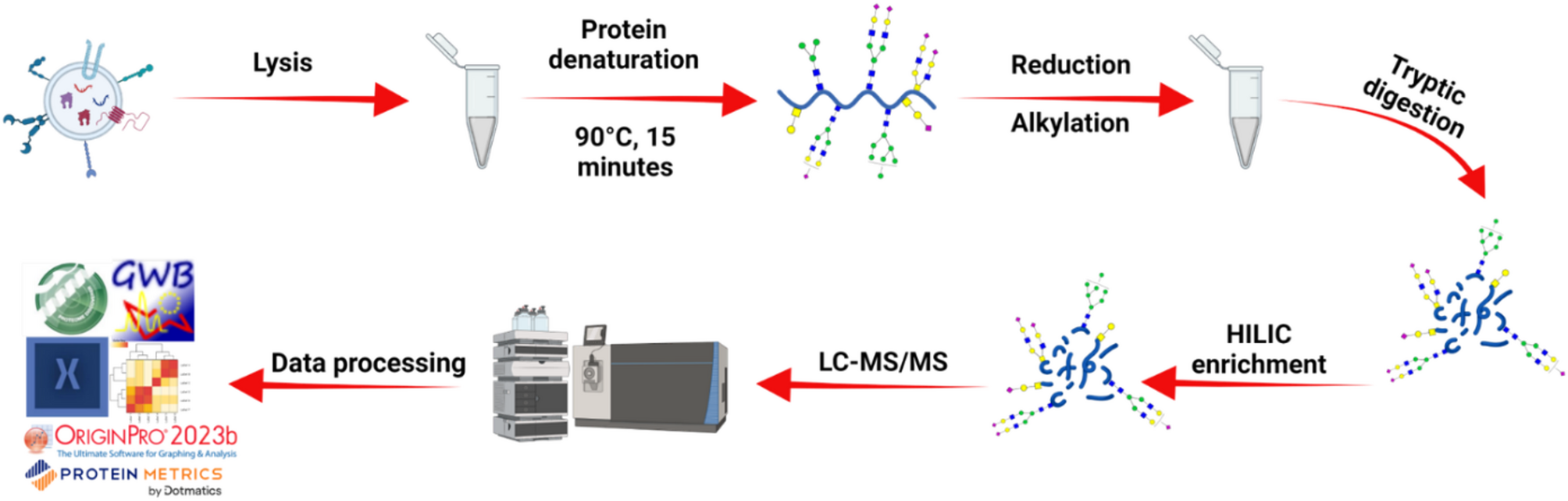
Isolated sEVs were lysed using a 5% SDC detergent solution with bead beating and ultrasonication. The released proteins were then denatured, reduced, alkylated, and digested with trypsin. Glycosylated peptides were subsequently enriched using HILIC and analyzed by LC–MS/MS.
They discovered that ultracentrifugation, especially with larger sample volumes, provides deeper insights into the glycan structures of the vesicles. However, when only a small amount of serum is available, the MagCapture kit still performs well. Importantly, they were the first to report site-specific glycan differences, called microheterogeneity, on sEV proteins from human serum, showing how complex and informative these sugar modifications can be.
Their work offers practical advice to researchers choosing between isolation methods and helps advance our understanding of how glycosylation shapes the biology of extracellular vesicles.


