Exosome RNA Exosome RNA Research & Industry News

Ductal carcinoma in situ, or DCIS, is considered a non invasive form of breast cancer. Although the abnormal cells are confined to the milk ducts, DCIS increases the lifetime risk of developing invasive breast cancer. In rare cases, patients even develop distant metastases without obvious local invasion. This raises an important question: can cancer related signals spread through the body before tumors visibly invade surrounding tissue?
Researchers at the University of Oklahoma Health Campus set out to explore this possibility by tracking tumor derived small extracellular vesicles, also known as exosomes, in an experimental model of DCIS.
What are small extracellular vesicles?
Small extracellular vesicles are tiny membrane bound particles released by cells. Cancer cells use these vesicles to send molecular messages, including proteins and RNA, to nearby and distant tissues. These signals can influence immune responses, metabolism, and even prepare distant organs for future metastasis.
While exosomes are known to promote tumor progression in invasive cancers, it has been unclear whether very early, non invasive lesions such as DCIS also release vesicles that travel throughout the body.
Tracking exosomes from early breast lesions
To answer this, the team used a mouse model in which human DCIS cells were introduced directly into the mammary ducts. The cancer cells were engineered to produce a light emitting tag attached to CD63, a well known exosome marker. This allowed the researchers to track vesicle release using bioluminescent imaging.
Over time, signals from tumor derived extracellular vesicles were detected not only in the mammary glands but also in distant organs. By day 14, vesicle signals were found in the spleen, liver, lungs, and brain. Later, they were also detected in the heart and in the bloodstream. Importantly, these signals appeared before there was clear microscopic evidence of invasive cancer.
This finding suggests that even when tumor cells remain confined within the ducts, they can still release exosomes that spread systemically.
Dissemination of MCF10DCIS CD63-NLuc sEVs
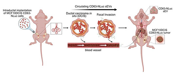
Graphical summary of the study investigating the biodistribution of ductal carcinoma in situ (DCIS)-derived small extracellular vesicles (sEVs) in a mouse intraductal model. Signals from DCIS cells expressing CD63-NanoLuc were detected in tissues and the circulation, correlated with enzymatic activity, and preceded histological invasion.
Effects on distant organs
The researchers also examined brain tissue using next generation RNA sequencing. By comparing gene expression at early and later time points, they found that immune, metabolic, and stress response pathways were reduced at 28 days after tumor implantation.
These changes indicate that tumor derived vesicles may influence distant organs at the molecular level, potentially conditioning tissues in ways that could support future tumor spread.
Why is this important?
The idea that preinvasive breast lesions can release extracellular vesicles into the circulation has important implications. It suggests that communication between early tumors and distant organs may begin much earlier than previously appreciated.
Understanding the timing and distribution of exosomes in DCIS could help explain how some patients develop metastases without clear local invasion. It also raises the possibility that circulating vesicles could serve as early biomarkers or even therapeutic targets in breast cancer progression.
By visualizing vesicle release in real time, this work provides new insight into how early stage cancer cells interact with the rest of the body long before tumors become overtly invasive.