 Exosome RNA Exosome RNA Research & Industry News

Diagnosing neurodegenerative diseases can be difficult because many disorders affect the brain in similar ways. Two conditions that are often challenging to distinguish are Lewy body disease (LBD) and Alzheimer’s disease (AD). Although they involve different biological mechanisms, their symptoms can overlap, and traditional blood biomarkers do not always clearly separate the two.
Researchers are now exploring whether extracellular vesicles, small particles released by cells, can provide new clues. These vesicles carry proteins, RNA, and other molecules from the cells that produced them. Importantly, extracellular vesicles from brain cells can cross the blood brain barrier and enter the bloodstream, meaning a simple blood sample may contain valuable information about what is happening inside the brain.
A research team at the University of Pennsylvania developed a new microfluidic technology designed to isolate specific types of extracellular vesicles from blood plasma. Their platform, called multiplexed Track Etch magnetic NanoPOre (mTENPO), uses magnetic labeling and tiny pores within a microfluidic device to capture vesicles originating from particular brain cell types.
Outline of the study and differential classifications performed
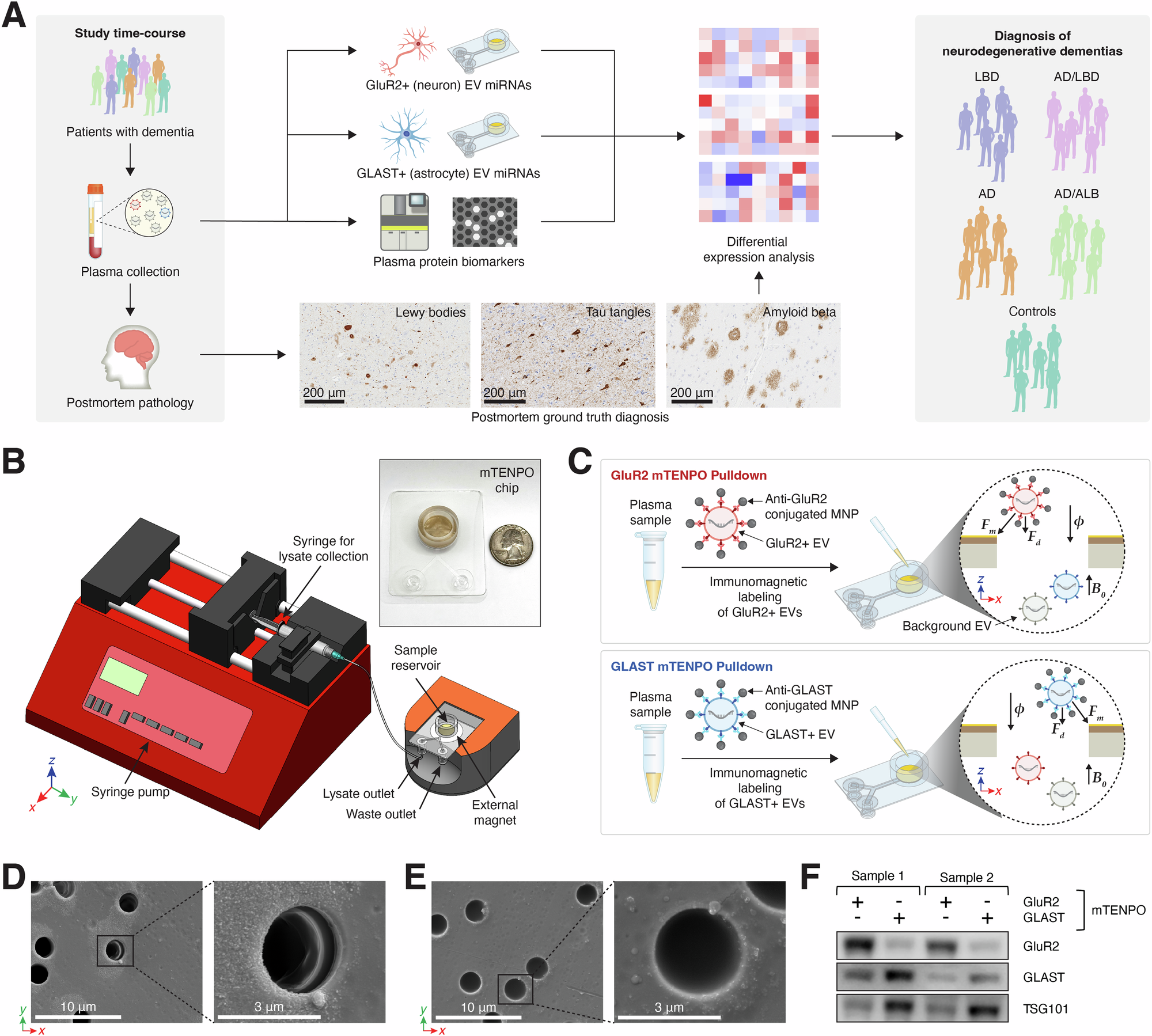
A Outline of study time-course and sample processing for EV isolation. Antemortem plasma samples with postmortem pathological confirmation of neurological diagnoses were processed to isolate cell-specific EVs using our mTENPO microfluidic platform, alongside plasma protein biomarkers using commercial digital ELISA, for patients with LBD (n = 30), AD (n = 31), AD/LBD (n = 30), AD/ALB (n = 19), and controls (n = 27). B The mTENPO platform, illustrating the external magnet, inlet reservoir, outlet ports, and tubing connections to syringe pumps. Syringes are connected to the waste outlet for blocking, washing, and sample addition steps, and then replaced and switched to the lysate outlet before captured EVs are lysed on-chip. The inset shows a photo of the mTENPO chip with a quarter for scale. C Schematic of operation of the mTENPO platform for cell-specific EV isolation using antibody-labeled magnetic nanoparticles (MNPs) for GluR2+ (top) and GLAST+ (bottom) EV pulldowns. D Scanning electron microscopy (SEM) images of GluR2+ EVs immobilized on the edges of pores of the mTENPO device’s surface. E SEM images of GLAST+ EVs immobilized on the edges of pores of the mTENPO device’s surface. F Representative cropped western blot images showing protein expression of GluR2, GLAST, and EV-associated marker TSG101 using mTENPO-isolated GluR2+ or GLAST + EV lysates from n = 2 human plasma samples.
The system allowed the researchers to independently isolate extracellular vesicles from neurons and astrocytes, two key cell types in the brain. Neuron derived vesicles were identified using the marker GluR2, while astrocyte derived vesicles were captured using the marker GLAST.
The team analyzed blood samples from 137 individuals whose diagnoses were confirmed by autopsy. The group included people with Lewy body disease, Alzheimer’s disease, mixed pathology, and control subjects. After isolating the cell specific extracellular vesicles, the researchers examined the small RNA molecules carried within them using miRNA sequencing.
These data were then combined with measurements of several well known Alzheimer’s related blood biomarkers, including amyloid beta 40, amyloid beta 42, tau, phosphorylated tau 181, and phosphorylated tau 231.
By integrating all of these signals, the researchers created a 15 feature biomarker panel that reflects multiple aspects of brain pathology. When the panel was tested using statistical validation methods designed to avoid overfitting, it achieved 95 percent accuracy in distinguishing Lewy body disease from Alzheimer’s disease. The model also showed a very strong diagnostic performance with an area under the curve of 0.96.
These findings suggest that analyzing cell specific extracellular vesicles from blood could significantly improve the ability to differentiate neurodegenerative diseases. Because extracellular vesicles carry molecular information directly from neurons and other brain cells, they may provide a clearer picture of disease processes than traditional blood biomarkers alone.
If further validated, technologies like the mTENPO platform could help move the field closer to noninvasive blood tests for neurological diseases, improving diagnosis and guiding treatment decisions for patients with complex neurodegenerative conditions.


