Exosome RNA Exosome RNA Research & Industry News

A new hypothesis proposed by researchers at Yale University suggests that extracellular vesicles (EVs), small particles released by cells, may not act alone in promoting healing. Instead, they may work by triggering a secondary response from nearby cells. When EVs are taken up by target cells, often macrophages, those cells then produce secondary EVs that carry out the actual biological effects, such as suppressing immune responses or promoting tissue repair.
In one example, EVs produced by CD8+ suppressor T cells activate macrophages to release secondary EVs that help calm overactive immune responses. In another case involving spinal cord injury, EVs derived from mesenchymal stromal cells (MSCs) interact with M2-type macrophages, leading those macrophages to produce EVs that aid in repairing damaged blood vessels.
This idea may help explain why MSC-derived EV therapies show promise across a wide range of diseases and tissue types, even when there is no perfect tissue match. The key may lie in the secondary EVs, which are produced locally and tailored to the specific environment or injury. These secondary responses could be the reason why EV treatments appear to have such targeted and disease-specific effects.
Circuits of primary administered EVs that activate new secondary EVs in the recipients

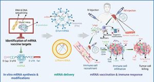
In some instances, when primary EVs act on targeted cells, they are stimulated by the transferred miRNAs or other cargoes to generate new secondary EVs. This usually features the involvement of the targeted cell DNA in generating secondary EVs that are then secreted to act on the final target cells to mediate the principal in vivo biological activity, illustrated by the three instances given here as examples (lower-right rectangles). First, one instance involves suppressor CD8+ T cell-derived primary EVs stimulated by high antigen dose immunologic tolerogenesis. These then act on local macrophages to induce their generation of secondary EVs, which then influence the final targeted Th1 effector T cells of DTH. Then, these secondary EV-producing targeted cells alter their T cell activation and subsequent cytokine production to participate in tolerance-mediated inhibition of delayed-type hypersensitivity (DTH), including the suppression of released INF-γ. In the second example, primary EVs derived from MSCs can aggregate in the lung to form primary EVs, or these primary EVs injected intravenously, promote maturation of M2 macrophages at the site of spinal cord injury (SCI) for production of TGF-β. Consequently, there is primary EV activation of these M2 Macs to produce secondary EVs that then target the local microvasculature to produce TGF-β receptors. Furthermore, the local microendothelial cells are induced to produce mRNA and subsequent proteins of vascular adhesion molecules to enable healing of the disturbed vascular permeability of SCI. A third instance, by analogy, is that LNP mRNA vaccines acting like primary EVs are taken up by local macrophages at the site of injection to produce secondary EVs for their transfer of mRNAs and miRNAs to the final targeted distant antigen-presenting, T, and B cells to generate a vaccine-induced immune response.\
If this mechanism holds true across other biological systems, it could provide important insights into how to design more effective and precise EV-based therapies in the future.