 Exosome RNA Exosome RNA Research & Industry News

Justus Ndukaife, associate professor of electrical and computer engineering and Chancellor Faculty Fellow, and his team have developed next generation nanotweezers that better analyze extracellular vesicles and aid in unraveling the mysteries of how cells package molecules and interact with one another.
Nanosized extracellular vesicles (EVs), though they vary in size and molecular cargo composition, are an important means for cells to communicate with each other. A significant research opportunity involves analyzing EVs individually to discern their biological roles in diverse diseases as well as leverage them for next generation therapeutics.
Studying single, intact EVs often relies on trapping individual particles, but existing methods face significant limitations. For example, optical tweezers – an approach recognized by the 2018 Nobel Prize in Physics – use a tightly focused laser beam to trap microscopic objects. However, the process is slow, as particles must be captured sequentially, and it is difficult to ensure that a new particle is trapped for each measurement. These constraints severely limit throughput and scalability.
Working principle of the Interferometric Electrohydrodynamic Tweezers (IET) system
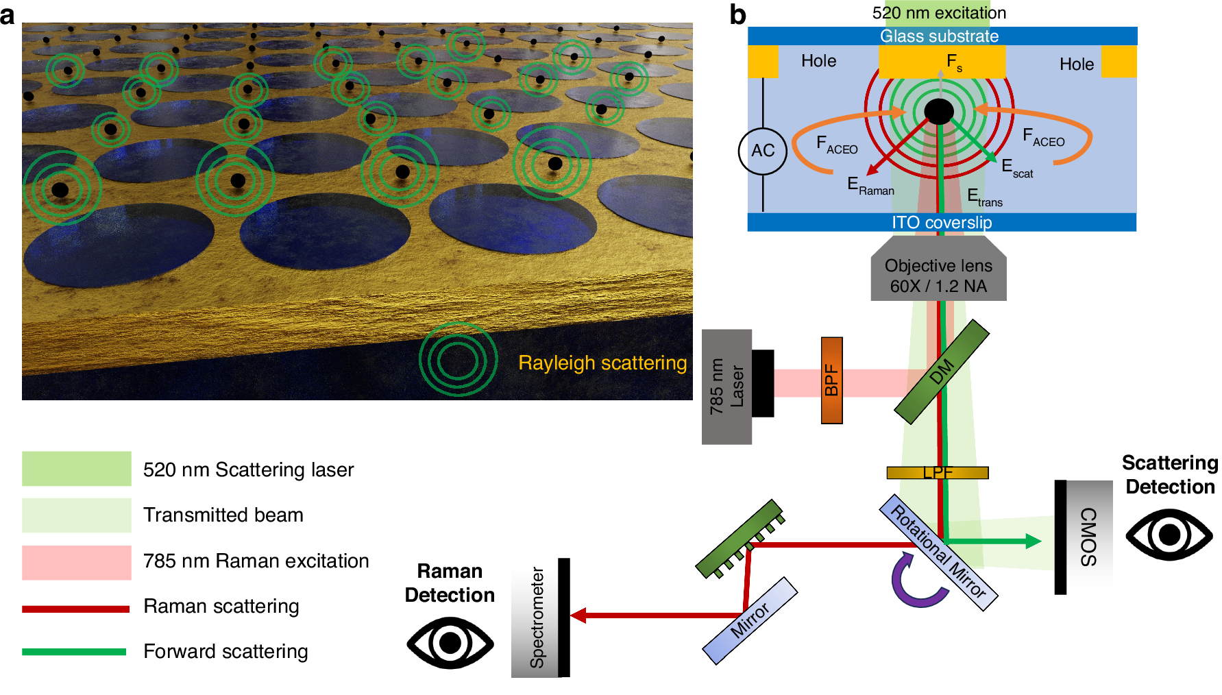
a Schematic view of the IET system capable of rapid parallel trapping of multiple particles and label-free interferometric imaging. b Illustration of the working principle and experimental setup of the IET system. Nanoparticles are rapidly transported by in-plane AC electro-osmotic (ACEO) flow and trapped at the nearest stagnation zones. Forces acting on the particles include FACEO, the drag force from ACEO flow, and Fs, the particle surface interaction force. Optical signals involved are Escat, the scattered electric field from the particle; ERaman, the Raman signal from the particle; and Etrans, the transmitted light after passing through the thin gold film. Optical components include: DM the dichroic mirror, BPF the band-pass filter, and LPF the long pass filter
To address this challenge, Ndukaife’s team has developed new technology – called interferometric electrohydrodynamic tweezers (IET) – that uses electrohydrodynamic flows to rapidly capture and trap thousands of nanoscale objects, including EVs and other nanoparticles, often within seconds. The platform integrates label-free interferometric imaging with Raman spectroscopy – a technique that involves illuminating a sample with a light source, typically a laser – enabling real-time characterization of individual particles without fluorescent labels or surface immobilization.
“Importantly, IET allows comprehensive analysis of nanoscale particles in their native, freely suspended state. By avoiding chemical staining or fixation, the technique minimizes measurement artifacts and preserves the intrinsic properties of each particle,” Ndukaife said. “Overall, the IET platform represents a significant advance in nanoscale particle trapping and characterization, opening new opportunities across nanomedicine, drug delivery, diagnostics, and environmental monitoring.”
Source – Vanderbilt University


