 Exosome RNA Exosome RNA Research & Industry News

Small extracellular vesicles, often called exosomes, have attracted major interest because they can carry proteins, RNAs, and lipids between cells and be harnessed for therapy. However, cells also contain many tiny vesicles that never leave the cell, and much less has been known about whether these intracellular vesicles could be useful for medical applications.
Researchers at the Tianjin Medical University Eye Hospital set out to explore this question by developing methods to isolate small intracellular vesicles from different cell types. They then directly compared these vesicles with well-studied extracellular vesicles to see how they differ in composition and function.
The researchers found that intracellular vesicles are generally smaller and can be obtained in higher quantities than extracellular vesicles. Importantly, they were taken up more efficiently by cells in both laboratory experiments and animal models. This suggests that intracellular vesicles may be especially effective at delivering therapeutic cargo into target tissues.
Therapeutic effects of MSC-sIVs and MSC-sEVs on blue-light-damaged mice (BL)
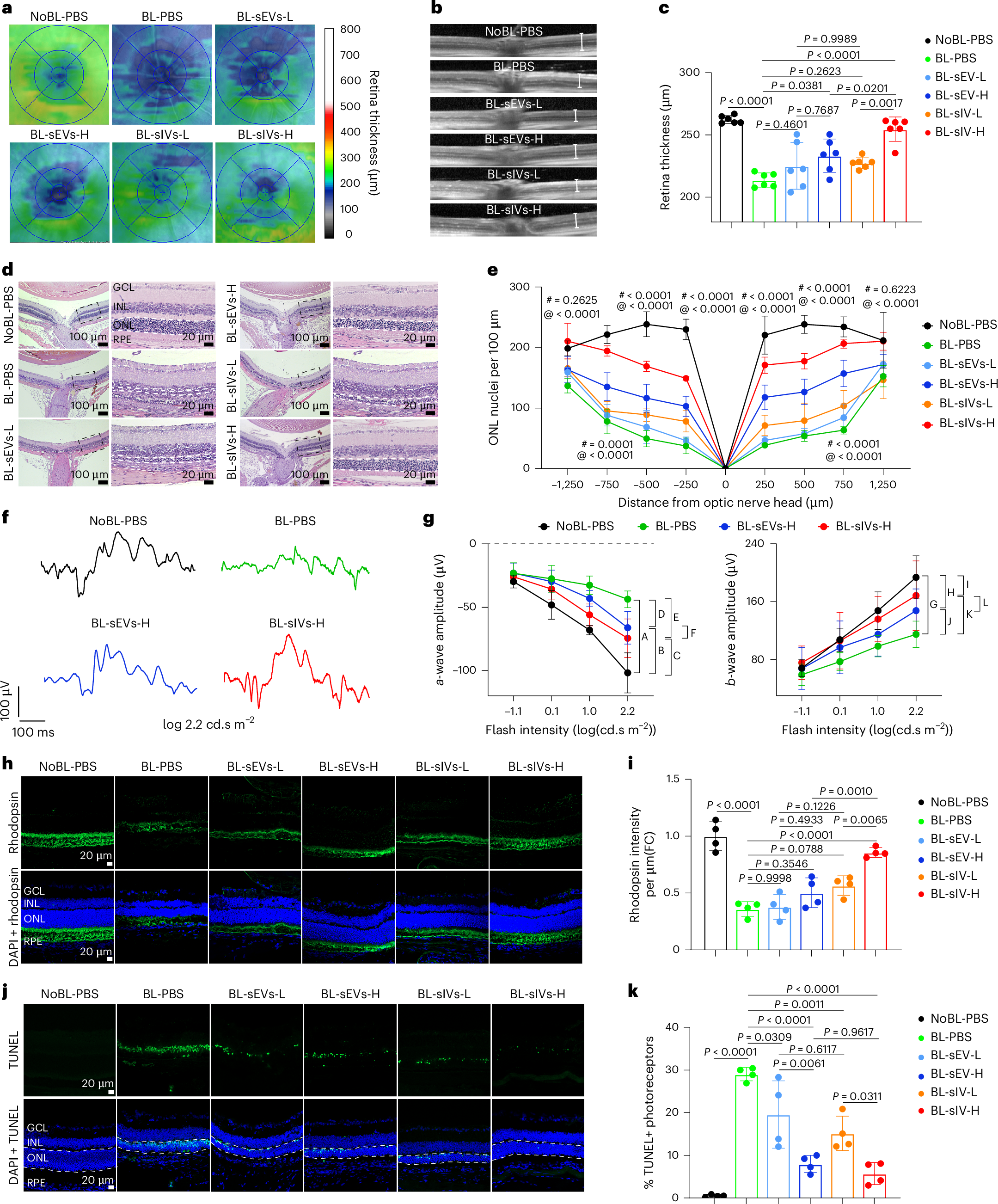
a, Representative topographic map of the retinal thickness. L, low-dose vesicles (2 μg); H, high-dose vesicles (4 μg). b, Representative spectral domain-OCT scans of the retina. White lines demarcate the region of interest for retinal thickness analysis. c, Retina thickness measurement. Six eyes, one from each mouse, were analysed (n = 6). d, Representative retinal images following H&E staining in each group (GCL, ganglion cell layer; INL, inner nuclear layer; ONL, outer nuclear layer). The number of nuclei within the ONL layer was quantified within the area delineated by the white dashed lines. e, The number of nuclei in the ONL extends outward along the dorsal/superior (positive values on the abscissa) and ventral periphery/inferior (negative numbers on the abscissa) directions, beginning at 250 μm from the optic nerve head and continuing every 250 or 500 μm. Four eyes, one from each mouse, were analysed (n = 4). ‘@’ indicates significant differences between BL-sIVs-H and BL-PBS, ‘#’ indicates significant differences between BL-sEVs-H and BL-PBS. Other statistical data are available in the source data. f, Representative ERG waveforms recorded in response to a stimulus flash (2.2 log(cd.s m−2)) from a single eye of a mouse in each treatment group. g, Average amplitudes of ERG a- and b-waves from each treatment group. Eight eyes, one from each mouse, were analysed (n = 8). The statistical differences between groups at a stimulation intensity of 2.2 were as follows: A < 0.0001, B = 0.0063, C = 0.2656, D = 0.0084, E = 0.0010, F = 0.4388, G = < 0.0001, H < 0.0001, I < 0.0001, J < 0.0001, K < 0.0001, L = 0.3081. Other statistical data are available in the source data. h, Immunofluorescence analysis of rhodopsin (green) expression in retinas. Blue, DAPI. i, Relative expression of rhodopsin in retinas. Four eyes, one from each mouse, were analysed (n = 4). j, Representative morphological images of retina sections. Green, TUNEL staining; blue, DAPI. White dashed lines indicate the area used for quantitative analysis. k, Ratio of TUNEL-positive nuclei in photoreceptors. Four eyes, one from each mouse, were analysed (n = 4). The data in c, e, g, i and k are expressed as mean ± s.e.m. and analysed using one-way ANOVA.
Molecular profiling revealed clear differences between the two vesicle types. Intracellular vesicles were enriched in proteins linked to the endoplasmic reticulum and Golgi apparatus, key compartments involved in protein processing and transport. They also carried distinct microRNA patterns associated with intracellular membrane systems and contained higher levels of certain phospholipids that support membrane structure and fusion.
To test their therapeutic potential, the team focused on vesicles derived from umbilical cord mesenchymal stem cells. In a model of retinal degeneration, intracellular vesicles reduced cellular stress in the endoplasmic reticulum and delivered neuroprotective factors more effectively than extracellular vesicles. These vesicles also showed a greater ability to load and deliver lipophilic drugs to retinal tissue.
Together, these findings suggest that intracellular vesicles are not just cellular byproducts but may represent a new and powerful class of delivery vehicles. By outperforming extracellular vesicles and exosomes in uptake, drug loading, and therapeutic impact, intracellular vesicles open new opportunities for treating diseases, particularly those affecting sensitive tissues like the retina.


