 Exosome RNA Exosome RNA Research & Industry News

Extracellular vesicles, or EVs, are tiny membrane-bound particles released by cells. They carry proteins, RNA, and other biologically active molecules that can influence nearby cells or travel to other locations, making them an important part of communication between cells.
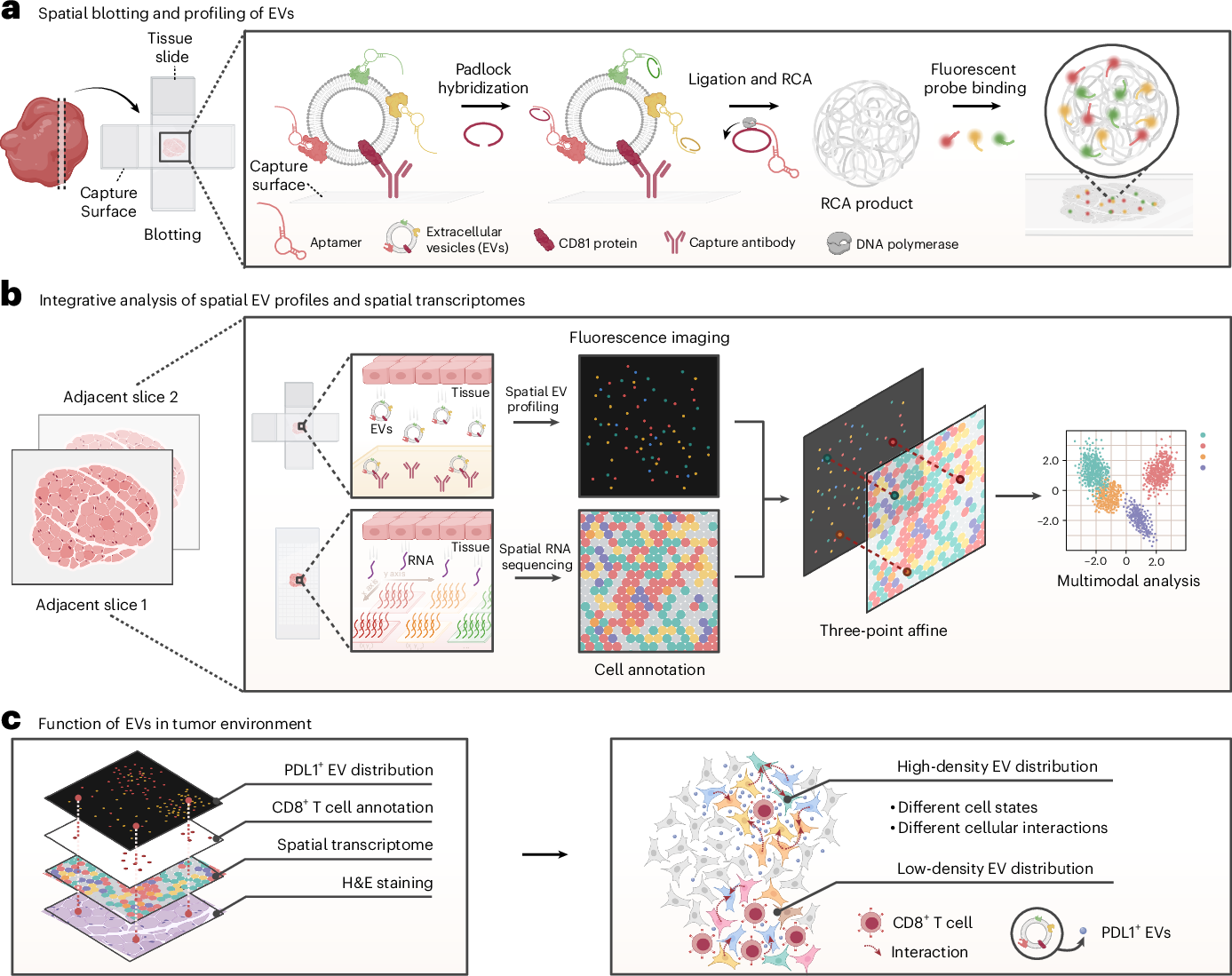
Researchers at Fujian Medical University have now described an approach called Spatial-EV-seq that makes it possible to examine where extracellular vesicles are located within tissues while preserving their spatial relationships.
Why the location of extracellular vesicles matters
Traditional methods for studying extracellular vesicles in tissues usually require the tissue to be broken apart. EVs can then be separated from other material using techniques such as differential centrifugation.
Although these methods provide valuable information about EV composition, disrupting the tissue removes information about where individual vesicles were originally located. Researchers can determine which EVs are present, but it becomes much harder to understand which cells released them, where they traveled, and which other cells they may be influencing.
This spatial information can be particularly important in diseases such as cancer, where neighboring regions of the same tumor can have very different biological characteristics.
Bringing spatial analysis to extracellular vesicles
Spatial transcriptomics has provided researchers with a way to measure gene activity while retaining information about where that activity occurs within a tissue.
Spatial-EV-seq applies a similar concept to extracellular vesicles. Instead of removing EVs from their tissue environment before analysis, the technique maps them while preserving information about their original locations.
The resulting EV maps can then be integrated with spatial transcriptomics data. Researchers can therefore compare the location of extracellular vesicles with gene expression patterns occurring in the surrounding cells.
This combination provides two complementary views of tissue biology, what cells are expressing and which extracellular vesicles are actually present in different regions.
Mapping extracellular vesicles in breast cancer
The researchers applied Spatial-EV-seq in a mouse model of breast cancer treated with an anti-PD1 immunotherapy.
They focused on extracellular vesicles carrying PDL1, a protein involved in suppressing immune responses. PDL1 can interact with the PD1 receptor on T cells and reduce their ability to attack cancer cells.
Spatial-EV-seq revealed distinct regions within tumors containing different levels of PDL1-positive extracellular vesicles.
Areas enriched in PDL1-positive EVs were associated with exhausted CD8-positive T cells and greater immunosuppression. In contrast, regions containing fewer PDL1-positive EVs maintained stronger immune activity.
These patterns suggest that the distribution of extracellular vesicles within a tumor could help shape localized immune environments.
EV location cannot always be predicted from gene expression
One particularly important finding came from comparing PDL1-positive EVs with expression of CD274, the gene that encodes PDL1.
Some tumor regions contained high concentrations of PDL1-positive extracellular vesicles even though nearby cells showed relatively low CD274 expression.
This mismatch suggests that the vesicles were not necessarily produced by cells at the location where they were detected. Instead, PDL1-positive EVs may have traveled from other parts of the tumor.
This distinction highlights an important limitation of using cellular gene expression as a substitute for directly measuring extracellular vesicles. A gene expression map can indicate where cells are producing particular molecules, but it cannot necessarily reveal where EVs carrying those molecules eventually travel.
Building maps of EV communication
The ability to directly visualize extracellular vesicles within intact tissue could provide researchers with a new way to investigate communication between cells.
In tumors, for example, Spatial-EV-seq could help determine where cancer-derived EVs accumulate and how their presence relates to immune cells, stromal cells, blood vessels, and other components of the tumor microenvironment.
Integrating these maps with spatial transcriptomics could also help researchers distinguish signals produced locally from biological messages transported into an area by extracellular vesicles.
Spatial-EV-seq therefore adds an important spatial dimension to EV research. By preserving the native distribution of extracellular vesicles, researchers can investigate not only what EVs contain, but also where they travel and how their location relates to cellular behavior within complex tissues.


