 Exosome RNA Exosome RNA Research & Industry News

When cells die, it’s not the silent end we might imagine. Researchers from La Trobe University have discovered that dying cells leave behind a unique structure that helps communicate with neighboring cells, even after death. This discovery adds an unexpected new layer to how our bodies manage cell turnover and immune response.
When a cell undergoes apoptosis, or programmed cell death, it typically sends chemical signals or releases tiny bubble-like particles called extracellular vesicles (EVs) to alert immune cells to clean up the remains. But the researchers found something new—an anchored, actin-rich structure left behind on the surface where the cell died. They named this the “Footprint of Death,” or FOOD.
Apoptotic adherent cells generate a membranous ‘footprint of death’ during cell retraction
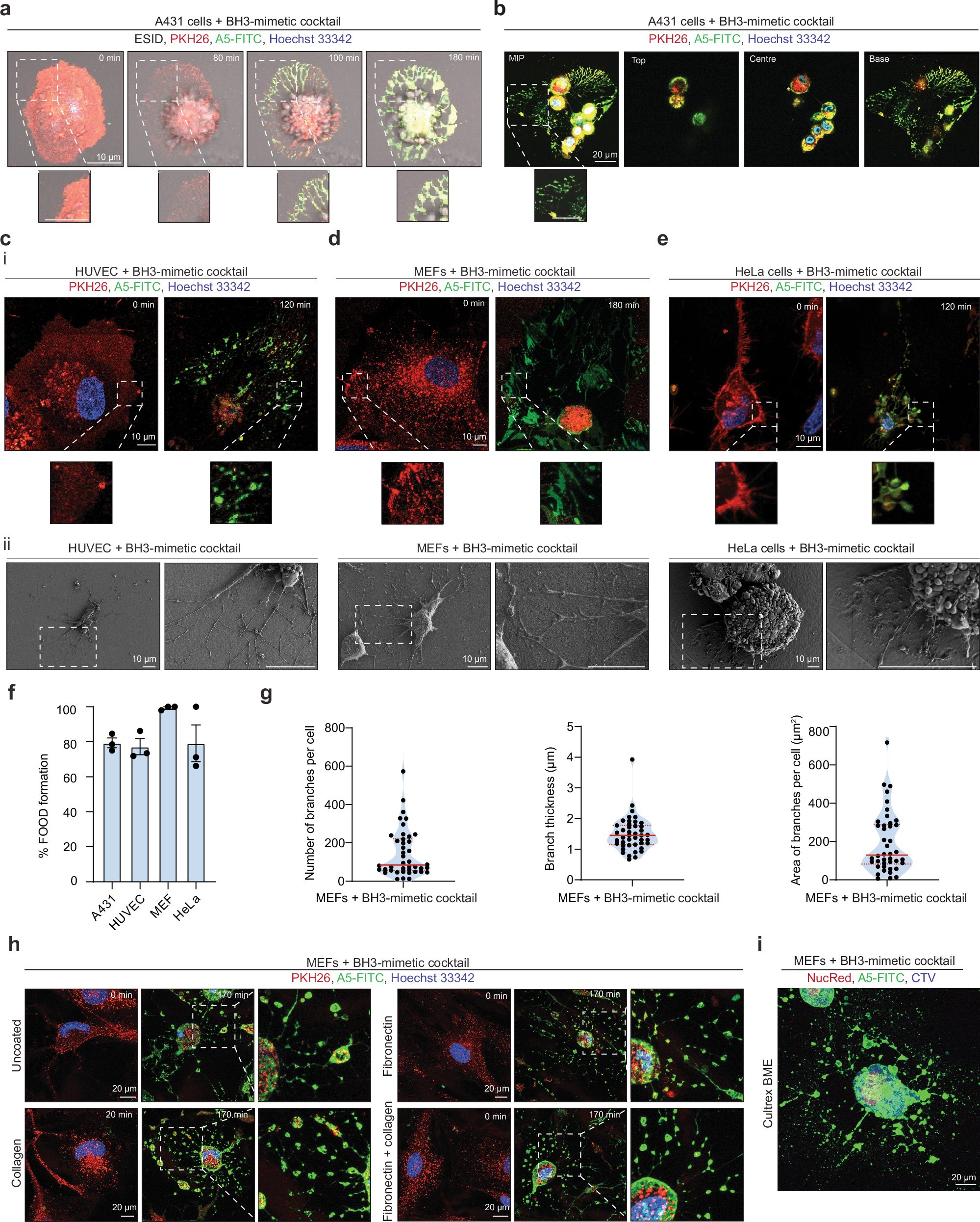
a, b A431 epithelial cells were treated with a BH3-mimetic cocktail (2.5 µM ABT-737, 0.5 µM S63845) and imaged by time-lapse confocal laser scanning microscopy (CLSM). Cell membrane and nucleus were visualised by PHK26 (red) and Hoechst 33342 (blue) staining, respectively, and exposed phosphatidylserine (PtdSer) was examined using A5-FITC (green). Cells in (b) are representative of cells 180 min post apoptosis induction. Maximum intensity projection (MIP) shown (left) and Z-stack images of top, centre and base of cells shown (left to right). Images demonstrating morphological changes following apoptosis induction in (c) HUVEC (2.5 µM ABT-737, 0.5 µM S63845), d MEF (5 µM ABT-737, 10 µM S63845), and e HeLa cells (2.5 µM ABT-737, 0.5 µM S63845) following treatment with a BH3-mimetic cocktail. Representative CLSM i (n = 3) and scanning electron microscopy (SEM) ii (n = 2). f Quantification of the percentage of apoptotic A431, HUVEC, MEF, and HeLa cells that form FOOD during the progression of apoptosis. Data is pooled from (n = 3) independent experiments. Apoptotic cells were identified based on A5 staining and apoptotic morphologies (i.e. cell retraction and membrane blebbing). Error bars represent s.e.m. g Quantitative analysis of the FOotprint Of Death (FOOD) generated by MEFs: number of branches per cell, branch thickness (μm), and area of branches per cell (μm2). Data is pooled from (n = 3) independent experiments. h Representative time-lapse CLSM MIP of MEFs on uncoated, collagen, fibronectin, and fibronectin and collagen coated chamber slides, treated with a BH3-mimetic cocktail. i Representative CLSM MIP of MEF in a Cultrex BME 3D-matrix. Cell cytosol and nucleus were visualised by cell trace violet (blue) and NucRed staining, respectively, and PtdSer exposed was examined using A5-FITC. At least three independent experiments were performed for all experiments unless otherwise specified.
These “footprints” aren’t just debris; they’re active communication hubs. Over time, they break apart into large extracellular vesicles that display phosphatidylserine, a well-known “eat-me” signal that attracts phagocytes (the body’s cleanup crew). Interestingly, under certain viral infection conditions, these structures can even carry viral particles, allowing infections to spread to nearby healthy cells.
The study reveals that FOOD formation depends on the protein ROCK1, which controls parts of the cell’s structure during death. This discovery changes our understanding of how dying cells communicate and interact with their surroundings—and highlights that even in death, cells can influence tissue health, immune function, and potentially disease progression.


