 Exosome RNA Exosome RNA Research & Industry News

Extracellular vesicles, or EVs, are tiny membrane-bound particles released by cells. They carry proteins, RNA, lipids, and other biological molecules that can provide information about the cells from which they originated. Because cancer cells also release EVs into the bloodstream, researchers are investigating whether these particles could serve as biomarkers for detecting and monitoring cancer.
A team led by researchers at Universiti Malaya have developed a new electrochemical biosensor designed to detect very small concentrations of extracellular vesicles in biological samples. The sensor combines an EV-recognizing aptamer with several nanomaterials to improve its sensitivity.
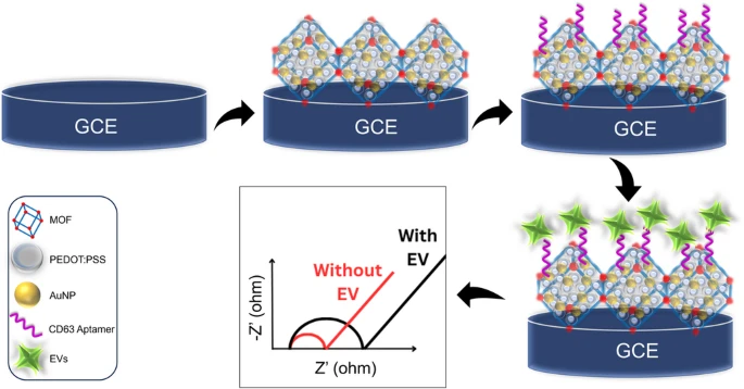
Why detecting extracellular vesicles is challenging
Liquid biopsy approaches aim to detect signs of disease in samples such as blood rather than relying exclusively on tissue biopsies. Extracellular vesicles are promising candidates because tumor-derived EVs can circulate through the body while carrying molecular characteristics associated with their cells of origin.
One difficulty is abundance. Tumor-derived EVs can be present at relatively low concentrations within complex biological fluids containing proteins, cells, lipoproteins, and many other particles.
A useful EV biosensor therefore needs to recognize the desired vesicles while producing a measurable signal even when relatively few particles are present.
Using an aptamer to recognize EVs
The researchers designed their biosensor around CD63, a membrane protein commonly associated with extracellular vesicles and frequently used as an EV marker.
Instead of using an antibody to recognize CD63, the researchers used an aptamer. Aptamers are short strands of DNA or RNA that can fold into three-dimensional structures and bind selectively to particular molecular targets.
The CD63 aptamer was attached to the surface of an electrode, allowing extracellular vesicles carrying CD63 to interact with the sensing platform.
Importantly, the sensor is label-free. This means that EV detection does not require the vesicles to first be tagged with fluorescent molecules, enzymes, or other labels.
Combining three materials to increase sensitivity
At the center of the biosensor is a glassy carbon electrode modified with a nanocomposite containing three different materials.
The first is a metal-organic framework, or MOF. These materials have highly porous structures that provide a large surface area and numerous sites where molecules can interact with the sensor.
The second component is PEDOT:PSS, a conductive polymer. Its electrical properties help transfer signals generated at the electrode surface.
The third component is gold nanoparticles. Gold nanoparticles are widely used in biosensors because they provide useful electrical properties and surfaces that can be modified with biological recognition molecules.
Combining the MOF, conductive polymer, and gold nanoparticles allowed the researchers to take advantage of the different properties of all three materials within a single sensing platform.
Detecting extremely low concentrations of EVs
Under optimized conditions, the biosensor achieved a limit of detection of 3.2 particles per microliter.
This level of sensitivity is important because disease-associated extracellular vesicles may be present at low concentrations, particularly when researchers are attempting to detect tumor-derived vesicles against a complex background of other biological material.
The researchers report that the sensor could reliably detect tumor-derived EVs at concentrations as low as approximately 10 particles per microliter in complex biological samples.
The system also demonstrated good reproducibility, selectivity, and storage stability, characteristics that will be important if EV biosensors are eventually adapted for routine laboratory testing.
Testing extracellular vesicles in serum
A biosensor that performs well under controlled laboratory conditions must also work in biological samples where many other molecules could interfere with detection.
The researchers therefore evaluated the platform using serum samples. Recovery rates ranged from 87% to 100%, indicating that the sensor could detect extracellular vesicles within this more complicated biological environment.
The biosensor was also able to distinguish differences in EV profiles between samples from healthy individuals and cancer patients.
These results suggest that electrochemical measurement of extracellular vesicles could potentially contribute to minimally invasive approaches for cancer screening. However, larger clinical studies will be necessary to determine how accurately and reliably the approach can distinguish patients with cancer from healthy individuals in real-world populations.
Extracellular vesicles as liquid biopsy biomarkers
One reason extracellular vesicles are attracting attention in cancer research is that they can provide access to biological information from cells that may otherwise require an invasive tissue biopsy to examine.
EVs circulate through biological fluids and can carry proteins and nucleic acids associated with the cells that released them. Detecting the vesicles is therefore one step toward accessing this molecular information.
Highly sensitive technologies could become particularly valuable when tumor-derived extracellular vesicles make up only a small fraction of the total EV population in a patient’s blood.
The new biosensor demonstrates how nanomaterials, electrochemistry, and molecular recognition can be combined to detect very small numbers of extracellular vesicles. Further validation in larger and more diverse clinical sample sets will be needed, but the approach provides another potential route toward incorporating EV analysis into minimally invasive cancer diagnostics.


