 Exosome RNA Exosome RNA Research & Industry News

A research team led by researchers at UCLA has developed a powerful new method to classify extracellular vesicles (EVs), tiny particles released by cells that carry genetic and protein information. These vesicles act like molecular messengers, reflecting the condition of the cells they come from. Because they can be collected from fluids such as blood or urine, EVs are being explored as a promising tool for non-invasive cancer diagnostics.
The challenge, however, has been accurately distinguishing between EVs released from healthy and cancerous cells. To solve this, the team used a technology called nanoaperture optical tweezers (NOTs), which can trap and analyze single vesicles using light instead of chemical labels. The light scattering patterns from each EV are then processed using deep learning, an advanced form of artificial intelligence.
Start-to-end deep learning-guided optical trapping workflow for single-EV analysis
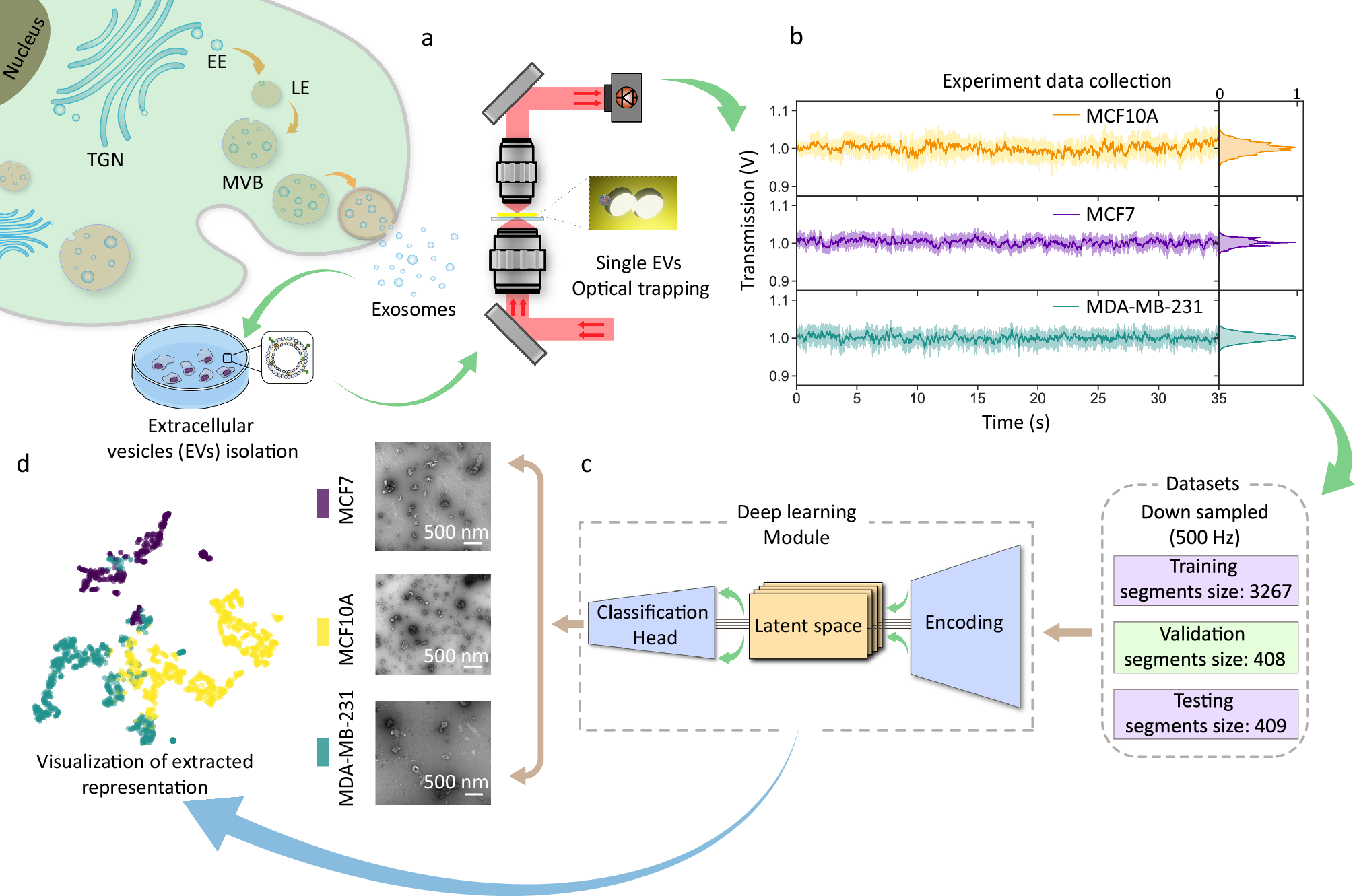
a Exosomes bud inward into MVBs and are released when MVBs fuse with the plasma membrane. (LE: intraluminal vesicles; MVBs: multivesicular bodies; EE: early endosome; TGN: trans-Golgi network). EV samples are diluted, placed on the sample stage, and covered by the double nanohole (DNH) aperture. Single EVs are trapped using a DNH optical tweezer, with trapping signals detected by a photodiode. b Representative trapping signals, their probability density functions, and power spectral density boxcar averages are shown, each fitted with a Lorentzian curve across different EV lines. c Signals are pre-processed and split (8:1:1) into training, validation, and test sets before being encoded and input into deep learning networks. The model is trained to optimize classification performance and extract latent features for visualization and downstream analysis. d Final outputs include EV class predictions and visualizations of the latent feature space.
The researchers designed a specialized neural network that includes both convolutional layers and a Kolmogorov–Arnold linear layer to recognize subtle differences in light signals. Their method achieved nearly perfect accuracy in classifying EVs from two types of cancer cells compared to those from non-cancerous cells.
This breakthrough shows the potential of combining cutting-edge optics and AI to create faster, more accurate diagnostic tools. Beyond cancer detection, the platform could help researchers profile EVs in real time, offering deeper insights into disease biology and paving the way for personalized medicine.


